|
||||||||||||||||||||||||||||
Gray Site
Teleoceras Rhinoceros
All magnifications refer to a 13 inch (32 cm) wide screen set to 800 x 600 pixels. The scanning resolution, directly proportional to magnification, is included as the last part of the file name. Right-click the image to obtain this information.
Earl Manning, Tulane University New Orleans, kindly provided the following ingformation, and the diagnoses used in the keys.
All of the material appears to derive from the forelimbs of a single adult Teleoceras individual, a grazing rhino ecologically more like a hippo than a modern rhino. Both unciforms show medial projections on their posterior processes. This suggests a late Hemphillian age (latest Miocene to earliest Pliocene) for the Gray Site Teleoceras, as it's a feature only common at the end of its lineage. It's part of an aborted attempt by Teleoceras to form a posterior articulation between the magnum and unciform, in order to stabilize the wrist (and attempt to fuse the two bones). The genus became extinct, along with other American rhinos, in the earliest Pliocene before the feature became well developed.
Harrison, J. A., and E. M. Manning, 1983. Extreme carpal variability in Teleoceras (Rhinocerotidae, Mammalia). Jour. Vert. Paleont., 3(1):58-64.
He also mentions the possibility of finding the remains of Aphelops, a taller browsing rhinoceros that lived in contemporaneous association with the grazing semi-aquatic Teleoceras.
See http://www.avph.hpg.ig.com.br/teleoceras.htm (Portuguese site) for a reconstruction of the animal in life.
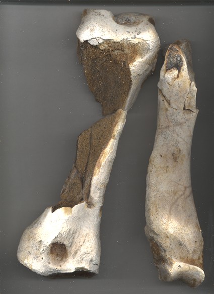
2/3 magnification. The bone on the left above was pictured on the front page of the Knoxville News Sentinel, uncredited. It is poorly preserved, and probably damaged by earthmoving activities. Parts of it turn to mush when wet. In a news item by reporter Fred Brown, Dr. Paul Parmalee of the University of Tennessee McClung Museum and Dr. Walter Klippel announced on September 18, 2000 that these had been identified as coming from Teleoceras, a small rhinoceros that lived in the Miocene Epoch. The identification was made by Dr. Michael R. Voorhies of the University of Nebraska, Lincoln http://www.unl.edu/geosciences/mrv/mrv.html who was involved in the discovery of the Ashfall fossil beds there. Earl Manning indicates these are two lower forelimb long bones, a partial right radius (proximal at bottom) on the left, and a partial right ulna (distal at bottom) on the right.
These, and all bones pictured below were recovered from blocks of disturbed weathered material dumped in a small area for a temporary road bed, and subsequently obliterated.
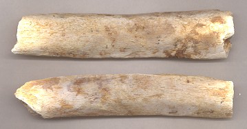
Sections of rib bone, 2/3X. Many short sections were recovered,
encased in a few broken blocks of stiff clay. These have a flat airfoil-
like cross-section.
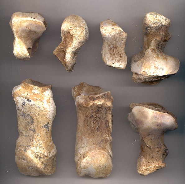
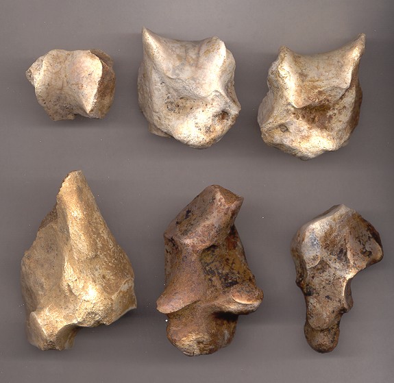
Carpals and metacarpals, natural size The two on the right are a left-right pair.
left magnum,
missing the post. process,
proximal viewright pisiform,
prox. at bottompartial left pisiform,
proximal at topright unciform,
distal viewleft metacarpal (MC 4),
anterior viewleft MC 2 in anterior view left unciform,
distal view
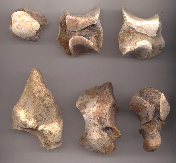
Opposite sides of bones in previous figure, natural size.
left magnum,
missing the post. process,
distal viewright pisiform,
prox. at toppartial left pisiform,
proximal at bottomright unciform,
proximal viewleft metacarpal (MC 4),
posterior viewleft MC 2 in posterior view left unciform,
proximal view
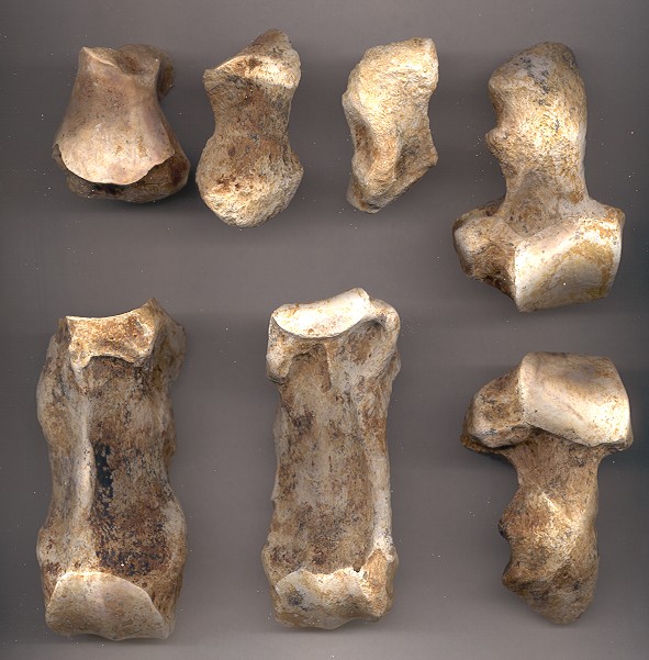
Additional carpal bones, natural size. The two on the upper right are a left-right pair.
trapezium,
proximal viewleft cuneiform,
lateral viewright cuneiform,
lateral viewprox. right MC 3
posterior viewright scaphoid,
distal viewright lunar,
lateral view
Opposite sides of bones in previous figure
trapezium,
distal viewleft cuneiform,
medial viewright cuneiform,
medial viewprox. right MC 3,
anterior viewright scaphoid,
medial viewright lunar,
medial view